Wednesday, July 12, 2017

Models should match observations

Ever since Galileo dropped two objects out of the Leaning Tower of Pisa (according to apocryphal accounts), it has been recognized that our scientific theories should be tested by observation. Sure, you could just make something up about how things work, but eventually someone will come along and just look to see if you're right.

Last week I wrote about an example in modern-day science where this hasn't happened. It was a small example...we are not talking about anything as momentous as testing the acceleration of gravity. But the example illustrated a point: sometimes ideas get so stuck in our heads that it may take a while before we realize that the observations are saying something different. 

Here is another example. Because of the action of the Dorsal nuclear concentration gradient (see here for more information about Dorsal), different genes are expressed in different locations around the circumference of the embryo. For example, the gene snail (sna) is expressed on the ventral-most 20% of the embryo (see figure below; by convention, the dorsal side is up and the ventral side is down). But measurements of where other genes are expressed were not so easy to do. Before we performed quantitative experiments in cross-sectioned embryos (where you could see the entire circumference of the embryo), it was thought that sog extended from about 25% to 70% around the embryo (see lavender colored arc on left figure). It was also thought that dpp was only in the cells on the dorsal-most 30% of the embryo.

Patterns of gene expression along the dorsal-ventral axis in the early Drosophila embryo (about 2.5 hours old). Illustration on the left (adapted from Stathopoulos and Levine, 2004) depicts sog as extending from 25% to 70% around the embryo (ventral-to-dorsal). dpp is shown as taking up the dorsal-most 30% of the embryo. More recently, fluorescent imaging in cross sections has become possible. The image in the middle is of a cross-sectioned embryo with several different genes detected by fluorescence. In particular, sog extends from 20% to 50% of the embryo circumference, while dpp takes up almost the entire dorsal-half of the embryo (adapted from Reeves and Stathopoulos, 2009). Both of these observations (plus those regarding other gene expression patterns) can be quantified and plotted as graphs (on right; adapted from Reeves et al., 2012). A border of a gene is defined as when it drops to 50% intensity.

However, after we began to make quantitative observations (using fluorescence) in cross-sectioned embryos, we could easily see that sog extends from 20% to 50% (see green fluorescence in middle figure and green curve in graph on right), while dpp takes up almost the entire dorsal half of the embryo (yellow fluorescence and yellow curve). However, even though this has been known since 2009, scientists are still publishing illustrations like the one on the left.

Wednesday, July 5, 2017

Shifting paradigms

In biology, as in all sciences, sometimes an idea gets so big, and so ingrained/entrenched in the culture, the original work does not need to be cited anymore. (Unfortunately, sometimes this means we often forget where the idea originally came from.) This idea becomes so pervasive, we believe it without even thinking about it. It becomes a fall-back idea, a foundation or bedrock, so to speak. Every new discovery is measured against it. Such an idea is called a paradigm.

However, sometimes, upon further review years later, we find that the original research that started the paradigm was flawed. Or the paradigm rests on a particular interpretation of the data from the original research, and not on the data themselves. In that case, the paradigm may shift. But because paradigms are so pervasive, it might take a lot of work and a long time for the shift to occur.

Now, to be clear, I am not claiming that any of the work I have been a part of constitutes a paradigm shift. Not at all. Usually, we reserve that moniker for truly momentous changes in an entire field of science, such as quantum mechanics or relativity. But some of our observations have shown that common illustrations of fly embryos, which have been in people's minds for decades, do have some inaccuracies.

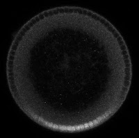
For example, consider the following illustration of a cross section of a fly embryo that is about 2.5 hrs old (left side of jpg below). The green represents the presence of the Dorsal protein (see here for more information about Dorsal). The common thought is that, as Dorsal enters the nuclei on the ventral side (bottom half of illustration), it depletes the surrounding cytoplasm of Dorsal. Hence, the cytoplasm around the ventral nuclei are dark. In contrast, Dorsal protein does not enter the nuclei on the dorsal side of the embryo (top half of illustration), so the nuclei are dark, but the surrounding cytoplasm is bright.


Illustrations like the one on the left have been around for decades. However, about eight years ago, we found out that this illustration is inaccurate. Yet, scientists are still drawing their embryos incorrectly today.

In theory, this "default view" of what happens with Dorsal should last only as long as it takes for someone to just look and see. In 2009, when I was a postdoc in Angela Stathopoulos's lab at Caltech, we published the first quantifiable (i.e., fluorescent) images of Dorsal-stained, cross-sectioned embryos (for example, see jpg above, right side). In several publications thereafter, we always saw the same thing: the cytoplasm is not bright on the dorsal side. In fact, there is no doubt there is just more total Dorsal protein on the ventral side. Yet, these illustrations of Dorsal in the embryo still persist today. I guess it just takes some time and effort for the "default view" to get out of our collective head.

Tuesday, June 27, 2017

The Happy Path and genome sizes

In software engineering, the Happy Path refers to the case when execution of the code happens with no problems or exceptions or possible errors. This scenario could also apply to other areas of engineering. For example, in chemical manufacturing, the unit operations are designed to give the desired output when the inputs are just as expected. But sometimes, disturbances upset the unit operation, so operating parameters like steam pressure, cooling water flow, etc., must be changed "on the fly" to make sure the product is up to specification. (That is what process control is for.)

The same principle is also true of other engineered systems. For example, while a car is designed to operate in a wide range of weather conditions, most of the time, you are on the Happy Path. But when it rains, it is a good thing the engineers built a subsystem of the car (wipers) to keep the water on your windshield from obscuring your view. An even more extreme case is your airbag. Hopefully, you'll never need to see your airbag in operation, but when you get off the Happy Path by crashing into something, you'll be glad the airbag is in place. Indeed, I am sure that careful inspection of the car's components, including the programming in the car's onboard computer, will show that a sizable fraction of the car is dedicated to situations that are not on the Happy Path.

In biology, "Happy Path" could be envisioned as when cells are grown under pristine laboratory conditions. If cells are kept at the just right conditions, with the just right amounts of nutrients, they will only execute the most basic sets of code and subroutines (the Happy Path). Under these cases, researchers have been able to strip down the genome to about 10% of its normal size. On the other hand, under wild (or uncontrolled) conditions, cells might face a myriad of challenges, and must execute various subroutines to proceed with growth/division. In this way, the vast majority of DNA code within the cell is there to ensure the cell continues to grow and divide, even when faced with unpredictable, suboptimal conditions.

Both the cell and man-made systems display this hallmark of complex, engineered systems: that a large fraction of the system's make-up is in place to provide robustness in the face of many disturbances.

Wednesday, June 8, 2016

Now on bioRxiv: A Facilitated Diffusion Mechanism Establishes the Drosophila Dorsal Gradient

We have recently put a manuscript on bioRxiv:

A Facilitated Diffusion Mechanism Establishes the Drosophila Dorsal Gradient http://biorxiv.org/content/early/2016/06/03/057091


In our manuscript, we have found that the Dorsal gradient is, at least in part, established by a facilitated diffusion mechanism. This is a way that the Dorsal protein can fight against its concentration gradient and accumulate on the ventral side of the embryo.  Without this, the Dorsal gradient likely would not get "tall" enough to specify all of the necessary genes, such as snail.

If you haven't checked out bioRxiv, it is the preprint server for biology hosted by Cold Spring Harbor Laboratory.  This is where researchers will post their findings before they get officially published in a peer-reviewed journal.

Thursday, January 14, 2016

How cells in a fruit fly embryo know what to do

Our favorite morphogen system is the dorsal-ventral (DV) axis specification in the Drosophila melanogaster (fruit fly) embryo.  In this system, a protein called "Dorsal" has a high concentration (in the nucleus) on the ventral side of the embryo and a low concentration on the dorsal side.  (By the way, if you're wondering why it seems the name is backwards -- why is Dorsal absent on the dorsal side, but present on the ventral side? -- you might want to read our post here.)  It also directs the cells along the DV axis to express different genes, in a concentration dependent fashion.  So Dorsal acts as a morphogen.

If you read our post introducing the idea of a morphogen, you know that the typical way a morphogen concentration gradient forms is by a small set of cells producing the protein, then diffusion causes the concentration of that protein to spread out.  This is not the case with Dorsal.  It is present everywhere, but cannot act because it's bound to an inhibitor protein called Cactus.  (Yes, seriously, it's called Cactus.  Again, you might want to read about why fruit fly gene names are so weird.)

But, on the ventral side of the embryo, a signal from the receptor Toll causes Cactus to be degraded, which releases Dorsal, allowing it to go into the nucleus and direct gene expression (see the above figure).  This "Dorsal nuclear concentration gradient" (see figure below) is one of the earliest signals to direct the cells in the fly embryo to differentiate into different types, specifying (from ventral to dorsal) muscle, neuron, skin, and an extraembryoinc tissue called the amnioserosa.  So the Dorsal gradient is very important, and is one of the more well-studied morphogen systems.

The Dorsal nuclear concentration gradient: This is a cross section of a 2.5 hr old fruit fly embryo that has been treated so that Dorsal protein fluoresces.  Dorsal protein is concentrated in the embryo's nuclei on the ventral side (bottom), and depleted on the dorsal side (top).

Wednesday, December 9, 2015

Of Course Biology Exhibits Engineering Principles

Just as every human-designed system must conform to standard engineering principles — discovered by trial and error — in order to work efficiently, so must biological systems at all levels be replete with engineering principles in order to maintain proper fitness.

Thursday, November 12, 2015

Why are fruit fly gene names so weird?

My lab studies the fruit fly (Drosophila melanogaster), which is one of the most well-studied animal systems.  Historically, fruit flies have been studied because the genetics is very easy.  Early on, the way to study genetics was to cause the flies to become mutated (altering their DNA randomly), and then see what happened.  Well, some pretty funky things happened.

Sometimes, the resulting flies looked all bristly -- researchers named that mutation "hedgehog".  Sometimes, they looked like a tube -- researchers called that mutation, well, "tube".  And so, names like "armadillo", "wingless", "cactus", "pipe", and "dachshund" were born.

Later, scientists found that these mutations mapped to genes within the flies' DNA.  Naturally, these genes were named after the mutation that the researchers originally found. 

It turns out that  one of the first mutations geneticists worked with was a spontaneous mutation that they named "white".  Well, guess what this mutation did?  It turned the turned the flies' eyes from red to white.  So when the genes were later named for the mutation involved in their discovery, the gene responsible for turning the flies' eyes red was called "white".  Brilliant!

So this is why many genes in the fly genome are named backwards.